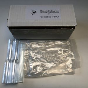
Description
This program is used to explore some of the major concepts and techniques that have revolutionized the biological sciences during the past two decades. Without the use of hazardous chemicals, students obtain an introduction and practical experience with the structure and isolation of DNA, cell fractionation, and gene cloning procedures. This three experiment program is set up for 16 students working in pairs and you may view the student manual attached below!
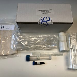
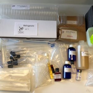
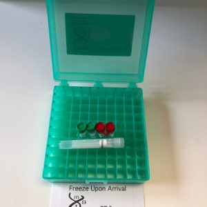
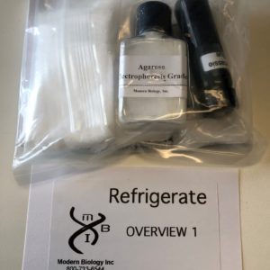
Basic Program 1 contains:
- * 19 Sterile Tubes
- 8 Tube Racks
- Nuclear Stain
- * 20 Petri Dishes
- 8 Glass Vials
- Sodium Dodecyl Sulfate
- * 24 Inoculating Loops
- 8 Glass Rods
- 2 Macropipetors
- Nuclear Buffer
- * Nutrient Agar + Ampicillin
- * 30 Sterile Transfer Pipets
- *Plasmid DNA
- Cheese Cloth
- * Nutrient Broth
- 10 Small Transfer Pipets
- Calf Thymus DNA
- * CaCl2
- DNAse I
- E. coli
- Calf Thymus Tissue
- Ampicillin
*Provided sterile and ready to use
Sample
A Sample from the Student Manual:
The laboratory manual is divided into two major sections. The first section provides basic information on the biology and chemistry of DNA. Students should be familiar with this material before advancing to the second section of the manual. The second part of the manual shows students how to apply what they have learned to perform three exercises in modem biology. The exercises deal with the structure function of DNA. Each experiment provides state-of-the-art information and each experiment can be completed within a 2 hour laboratory session. A background information section is given for each exercise and students should be required to read this material prior to performing the exercise in the laboratory. Study questions are found at the end of each exercise. Students derive answers to these questions by integrating the background material found in the text with the results of the experiments.
ParT a. background InformaTIon
I. Nucleic Acids: A Review of the Basics
The concept that chromosomal units known as genes transmit heritable information from parent to offspring was founded in the late 19th century. However, a description of genes in terms of their unique structural and functional properties is relatively new. We now know that genes are composed of a type of nucleic acid called deoxyribonucleic acid (DNA). The DNA molecule not only directs its own reproduction but also stores all the information that determines the types of proteins produced during the lifetime of an organism. In so doing, DNA orchestrates the complex reactions and structures characteristic of an organism and its offspring. Ribonucleic acid (RNA), the second major category of nucleic acids, is involved principally in the transmission of genetic information and in protein production. The structure and function of DNA and RNA can most easily be understood by examining the chemical composition of the nucleic acids.
Nucleotides – Building Blocks of Nucleic Acids
Under the proper conditions, nucleic acids can be broken down to low- molecular weight products of three types: a pentose (or 5 carbon) sugar; purines and pyrimidines; and phosphoric acid (Figure 1). The phosphate group is responsible for the strong negative charge of nucleic acids. The pentose sugar from RNA is always ribose and that from DNA is 2-deoxyribose. These sugars differ only by the presence or absence of a hydroxyl group on carbon 2 (so-called 2’). The numbers assigned to the five carbon atoms are shown in Figure 1. The purines and pyrimidines are often called nitrogenous bases (or, simply, bases). The major purine bases in DNA and RNA are adenine (A) and guanine (G), and the major pyrimidines in DNA are cytosine (C) and thymine (T). RNA contains the base uracil (U) in place of thymine. The sugars and phosphates are readily soluble in water. That is, they are hydrophilic. In contrast, the bases are hydrophobic in that they display limited solubility in water. As will be discussed below, these differences in water solubility are extremely important for the structure of the DNA molecule.
A nucleotide consists of a pentose sugar, a nitrogenous base and a phosphate group structured as shown below. The high-energy storage compound, adenosine triphosphate (ATP), is a well known nucleotide found in biological systems.
The Polynucleotide Chain
Nucleic acids are polynucleotides and have the general structure shown below:
A polynucleotide is composed of repeating nucleotide units linked into chains by phosphodiester bonds that join the 5’ carbon of one ribose or deoxyribose group to the 3’ carbon of the next sugar (Figure 2). The sequence or order of nucleotides in a polynucleotide chain is often abbreviated by a 1-letter code (e.g., G-C-A-T-A) with the 5’ end of the chain written at the left. A typical RNA molecule is a single- stranded polynucleotide chain. As will be described below, DNA usually contains two polynucleotide strands coiled around one another to form a double-stranded helix. The number of nucleotide units in a nucleic acid chain varies tremendously depending on the nucleic acid type. For example, each chromosome from a higher organism is thought to contain a single, very long DNA molecule. A DNA molecule from the largest human chromosome is composed of approximately 5.4 x 10⁸ nucleotides, which corresponds to a molecular weight of the order of 10 11 and a length of about 4cm. On the other hand, transfer RNA molecules generally contain only 70-80 nucleotides.
DNA Structure
In early physical studies of DNA, a variety of experiments indicated that DNA molecules occur in long helixes with each helix being formed from two or more polynucleotide chains bound side by side. Chemical analyses also demonstrated that the phosphate groups were on the outside of the helix and that the number of A and T residues in DNA were always equal, as were those of G and C. With these facts in mind, Watson and Crick in 1953 proposed that the DNA molecule actually consists of two polynucleotide chains coiled around the same axis to form a double helix (Figure 3). In this model, the hydrophilic sugar-phosphate groups follow the outer edges of the molecule where they can interact with water. The hydrophobic bases face inward toward each other in the molecule’s center and thus avoid contact with water. The two polynucleotide strands run in opposite directions (they are anti- parallel) and are held together primarily by hydrogen and hydrophobic bonding between the bases, where A is always paired with T, and G with C. These complementary bases have an affinity for each other such that, when they are paired, they contribute to the overall stability of the DNA helix. Because of this complementary base-pairing, the sequence of bases in one polynucleotide chain determines the sequence in the other. For example, if the bases along one strand are arranged in the order T-G-C-T-A-G, the opposite bases on the complementary strand will be A C-G-A-T-C. This fact is of extreme biological significance because it explains how a DNA helix in the chromosome directs the formation of copies of itself and directs the formation of RNA molecules with its specific informational content. The B-form DNA shown in Figure 3 is the most common of the DNA types. It is a right-handed double helix and contains about 10.5 nucleotide pairs per helical turn.
Biological Role of DNA and RNA
DNA is an information molecule with two general functions (Figure 4). First, DNA plays a central role in the propagation of the species and the determination of the heritable characteristics of the cell and its descendants. Prior to the time of each cell division, the two strands of the DNA helix separate from one another and each serves as a pattern or template for the synthesis of a new, complementary chain. This process of DNA biosynthesis is known as replication. One of the double helices formed is then transmitted to one daughter cell, and one to the other. Although the principle underlying DNA replication is straightforward, the actual mechanism responsible for the replication process in the cell involves an array of enzymes and regulatory proteins.
The informational content of DNA also determines the types of proteins that are produced by a cell. In this manner, the DNA molecule functions as a blueprint for all cellular processes that go on during the lifetime of an organism. In the first step along the information pathway from DNA to protein, a segment of DNA is copied into a complementary strand of messenger RNA (mRNA) by a process known as transcription. Transcription begins when an enzyme called RNA polymerase binds to a specific sequence on the DNA known as the promoter. At this site, the enzyme unwinds a small segment of double helix, exposing the bases of the two single strands of the DNA molecule. One of these strands is then transcribed. As the polymerase travels along the DNA, ribonucleotides with bases complementary to the DNA are added to the growing chain. For example, the DNA segment C-G- T-A-T G is transcribed into G-C-A-U-A-C in the mRNA. Each sequence of three nucleotides in the mRNA is called a “codon,” and codes for one amino acid. Since most polypeptide chains contain between 100 to 1,000 amino acids, an mRNA must be at least 300 to 3000 nucleotides long. Therefore, a gene that codes for a polypeptide chain must contain at least 300 to 3,000 base pairs.
The translation of mRNA into protein is a complex process that occurs on particles called ribosomes. This process requires ribosomal RNAs (rRNAs) and transfer RNAs (tRNAs). These RNA species do not specify proteins themselves but rather take part in decoding the information carried by the mRNAs. At one end of each tRNA molecule is a nucleotide triplet called the “anticodon,” which is complementary to an mRNA codon. A specific amino acid is bound to the opposite end of each tRNA molecule. On the ribosome, tRNAs carrying amino acids associate with the mRNA by way of complementary base-pairing at the anticodon-codon sequences. As the ribosome moves along the mRNA, the amino acids carried by the tRNAs are linked to the growing polypeptide chain. In this manner, the order of codons along the mRNA directs the amino acid sequence of a polypeptide chain. The translation process occurs on the surface of the ribosomes. These particles, which are composed of rRNA and ribosomal proteins, serve to bring together the mRNAs, the tRNA and other factors that are required for protein production.
Replication
The two DNA strands separate and each serves as a template for the synthesis of a new, complementary polynucleotide.
Transcription
One strand of the DNA serves as a template for the synthesis of complementary RNA.
Translation
The mRNA associates with ribosomes and its nucleotides are matched three at a time to a complementary set of three nucleotides in a specific tRNA molecule. The tRNA molecule carries an amino acid and when it associates with the mRNA, the amino acid is added to the growing polypeptide chain.
 Due to Customs restrictions, we only accept orders from educational institutions within the Continental United States, Alaska or Hawaii.