Due to Customs restrictions, we only accept orders from educational institutions within the Continental United States, Alaska or Hawaii.


Description
Microtubules are hollow cylinders made up of polymers of the protein tubulin. Microtubules are major components of cilia and flagella, which are tail like projections that are covered by a plasma membrane and extend outwards from the cell. Motile cilia are used for locomotion and food gathering by some protozoa and are found in the lining of the trachea, where their wave like motion propels mucus, dust and other foreign matter out of the lungs.
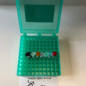
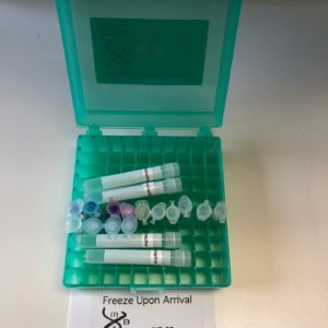
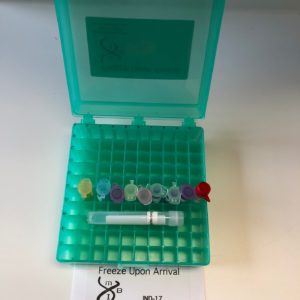
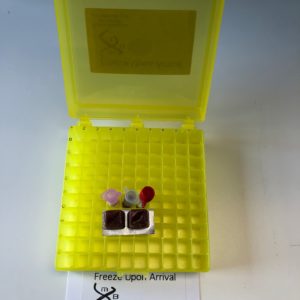
In this exercise, students use the powerful technique of immunohistochemistry to localize tubulin in the esophagus and trachea. The provided tissue sections are exposed to a tubulin monoclonal antibody, which binds to the tubulin. The sections are then incubated with a secondary antibody, which binds to the first antibody. The second antibody is complexed with peroxidase, which catalyzes a color producing reaction. Following the addition of a non-toxic peroxidase substrate, the peroxidase converts the colorless substrate to a blue product, thus “staining” the area containing tubulin. As can be seen in the photographs in panels A-C at the right, the cilia that line the trachea are stained in a highly selective manner in keeping with the high concentrations of microtubules that make up these structures. This exercise was designed for eight groups of students. 5 groups use the above procedure, one group carries out the procedure without the tubulin antibody (as an important negative control) and two groups stain their sections with hematoxylin and eosin as seen in panel D. The analysis is supplied complete with all of the instructions, chemicals, and accessories needed to perform perhaps the most important method used by the cell biologist. Sufficient antibodies, peroxidase substrate and other reagents are provided so that between 5-10 additional slides (provided by the instructor) can be analyzed by the procedure. Distilled water and ethyl alcohol or isopropyl alcohol are needed but not included.