Description
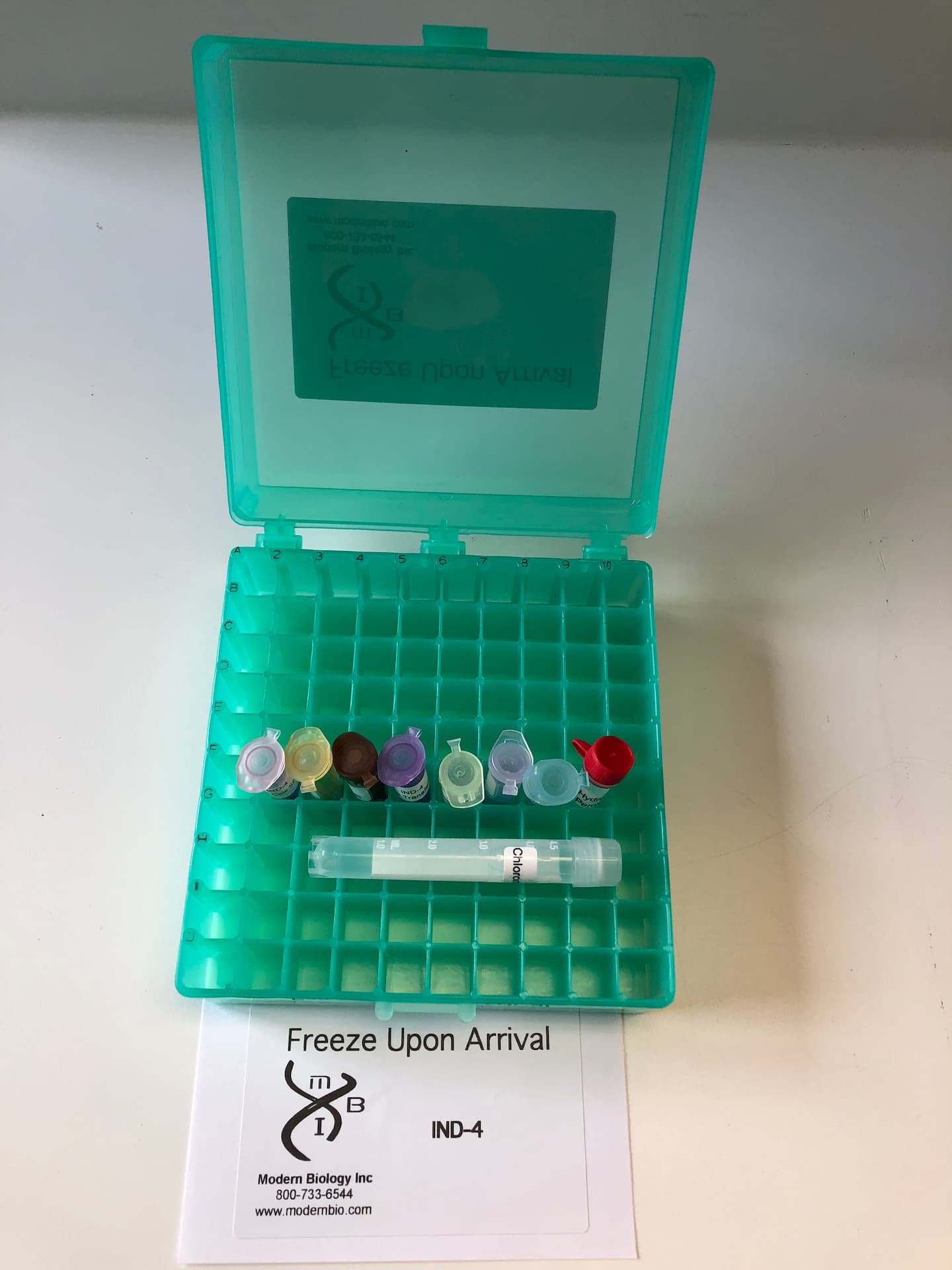
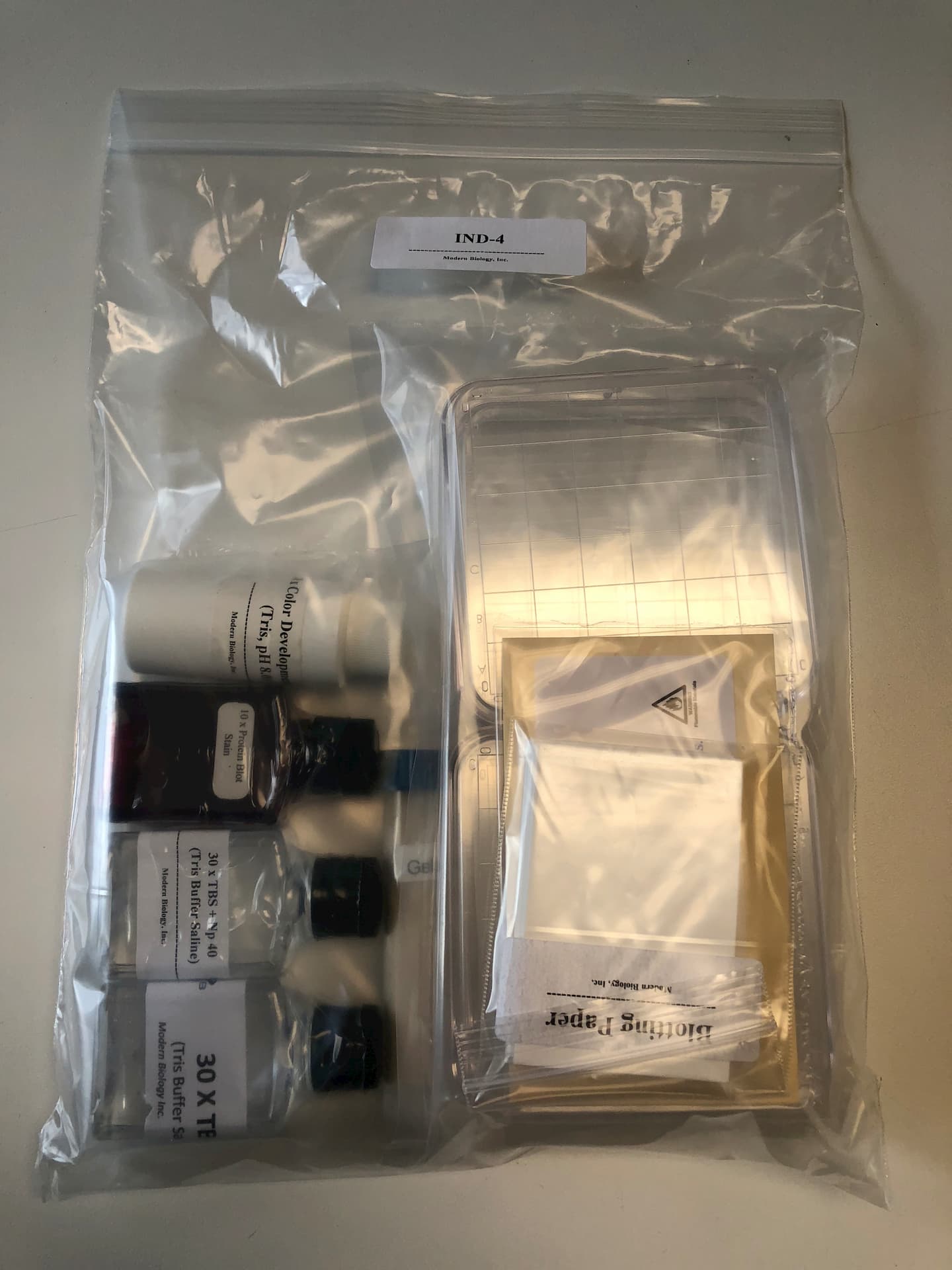
In most mammals, antibody production does not occur until after birth. The newborn calf receives antibodies in milk and these antibodies are responsible for passive immunity during early postnatal life. Thus, fetal calf serum is devoid of antibodies, neonatal calf serum contains antibodies of maternal origin while serum from the adult contains large amounts of antibodies made by the mature immune system. This fascinating developmental scheme is illustrated in this exercise. First, students electrophorese serum proteins from fetal, newborn, and adult animals on an agarose gel and the separated proteins are transferred onto a nitrocellulose membrane by the Western Press-Blot procedure shown on the facing page. All proteins on the blot are stained with protein blot stain and then the major class of circulating antibodies (IgG) is detected by using an immunological procedure. The results show that albumin and transferrin are present in sera from all stages while the antibodies are absent in fetal serum but present at high levels in serum from the adult. This analysis can be performed during a single 3-hour, or two 2-hour laboratory sessions. The exercise was designed for 8 groups of students and includes: fetal calf serum, newborn calf serum, adult cow serum, anti-Cow IgG (peroxidase linked), nitrocellulose, blotting paper, protein blot stain, Tris buffer saline (TBS), TBS + NP40, color development buffer, chloronapthol, hydrogen peroxide, dishes for blot incubation, and gelatin.
Sample
Background Information
I. Serum Proteins
Blood is a remarkable tissue containing cellular elements (erythrocytes, leukocytes and platelets) suspended in a liquid medium called plasma. Whole blood or plasma clots upon standing and if the clot is removed, the remaining straw colored fluid is called serum. Serum has basically the same composition as plasma except that it lacks certain proteins (fibrinogen and some clotting factors) that are involved in the clotting process.
Serum contains a variety of small molecular weight components as well as hundreds of different serum proteins. The serum proteins are usually divided into albumin and globulin classes. Serum albumin is the major protein found in serum. In humans, the albumin constitutes about 50% of total serum proteins. Albumin functions in the transport of many substances in blood and plays an important role in the control of plasma osmolarity. The serum globulins can be subdivided into three major fractions. These fractions are the alpha, beta, and gamma globulins. Each globulin fraction contains a large number of different proteins. For example, the protein transferrin is only one of the proteins found in the beta -globulin fraction. The antibodies are found in the gamma globulin fraction of serum. For this reason, the antibodies are also called immunoglobulins (abbreviated as Ig). In mammals, there are 5 major classes of immunoglobulins (IgA, IgD, IgE, IgG, and IgM). The major class of immunoglobulins in blood is IgG and this class constitutes about 10% of total serum proteins.
II. The Antibody Molecule
The immune system consists of a diverse set of cells, tissues and organs and about 1012 antibody molecules. The major function of the immune system is to protect the organism from viruses, bacteria, protozoans and larger parasites. When the organisms enter the vertebrate body, macromolecules on their surfaces induce the production of specific antibodies that appear in the serum of the infected animal. The antibodies, in turn, combine with these foreign macromolecules thereby rendering the invading organisms inactive and noninfective.
The macromolecules that elicit antibody production are called antigens and are most often proteinaceous in nature. Although antigens are frequently components of foreign organisms, purified foreign proteins will serve as antigens in that they will stimulate the formation of antibodies when injected into a suitable test animal such as the rabbit. Each antigen possesses features that are recognized by the antibody and these features constitute the antigenic determinants or epitopes. An antigenic determinant recognized by an antibody molecule may be a unique shape or a sequence of about 5 to 10 amino acid residues on the protein molecule. It follows that each protein possess a large number of potential antigenic determi¬nants and when a foreign protein is injected into an animal, antibodies to different determinants on the protein are produced and appear in the serum. This antibody containing serum is called an antiserum. Thus, an antiserum generated by immu¬nization with one purified protein usually contains a large number of different antibodies which recognize determinants along the protein.
The structure of the most common type of antibody is best represented as a Y-shaped molecule as shown in Figure 1. The molecule is composed of two identical light chains (L) and two identical heavy chains (H). Each L and H chain contains about 220 and 440 amino acid residues, respectively. The amino terminal ends of the L and H chains come together to form the antigen binding sites as shown in Figure 1. Two identical antigen binding sites are found on each antibody molecule. The portions of the L and H chains of a given antibody that form the antigen binding sites contain amino acid sequences which are different from all other antibodies. These so called hypervariable regions are responsible for the binding specificity of antibodies.
III. Development of the Immune System
Fetal and newborn mammals do not make antibodies. Thus, a source of antibodies is needed to combat infection during early postnatal life. In some mammals including humans, antibodies are transferred from the mothers blood through the placenta to the fetus. These maternal antibodies create temporary passive immunity which occurs in humans during the first few months of life. After this time, the newborn’s immune system begins to produce large amounts of antibodies and this condition persists into adulthood.
The first step in Western Blotting is to electrophoretically separate the serum protein samples in agarose gels. Next, the gel is placed against a specialized membrane made of nitrocellulose. The proteins are then forced out of the gel by applying gentle pressure and are trapped on the nitrocellulose membrane. As a result, a replica (blot) of the electrophoretically separated proteins is produced on the nitrocellulose membrane.
In the cow and related ungulates (horse, sheep, goat, etc.), antibodies are not transferred from the maternal to the fetal circulation because the large antibody molecules cannot cross the placental barrier in these animals. Thus, fetal serum in these species is devoid of antibodies. Antibodies appear in the serum of these animals only after the young have suckled and received the first milk. This milk, called colostrum, contains large amounts of antibodies which are responsible for the transmission of immunity from mother to young. Correspondingly, the newborn adsorbs these antibodies from the gastrointestinal tract which is permeable to large protein molecules (like antibodies) during the first few days after birth.
IV. The Western Press-Blot
1. Run agarose gel.
2. Build “sandwich” consist¬ing of gel casting tray, paper towel, blotting paper, gel, and nitrocellulose membrane.
3. Transfer separated proteins to nitrocellulose membrane by applying gentle pressure on the gel casting tray.
Antibodies are frequently used to identify specific proteins because they recognize unique antigenic determinants along a protein molecule. For example, human serum albumin injected into a rabbit makes the animal produce anti-human albumin antibodies. These antibodies are directed against determinants of human albumin (shapes and/or amino acid sequence) that are not found on the rabbit albumin molecule. When the anti-albumin serum is mixed with human albumin, the antibodies in the serum react strongly with the albumin and an antibody-antigen complex is formed. Similarly, the antiserum will react strongly with albumin isolated from gorilla, orangutan and baboon. In contrast, the anti-human antiserum will not react with transferrin or any other protein in serum.
In this experiment, you will be comparing serum proteins from various stages in the development of the cow. First, you will observe the patterns of electrophoreti¬cally separated proteins present in sera from fetal, newborn, and adult cow. You will then perform an immunological procedure known as Western Blotting. Through a series of steps (described below), this procedure enables the investigator to visualize proteins that react with a specific antiserum after the proteins have been separated by electrophoresis.
4. Detect all proteins on the membrane with protein blot stain.
5. Detect specific proteins on the nitrocellulose membrane with immunological techniques.
The next step is to incubate the membrane with antibodies which react with the proteins trapped on the membrane. In the present experiment, you will use antibodies that were generated in rabbits against cow IgG. These antibodies will bind to the membrane-trapped IgG and not to other proteins in the sera samples. Gelatin is added to the membrane before and during antibody incubation in order to minimize non-specific protein-antibody interaction.
Since the antigen-antibody complexes are not colored, they must be treated in some way in order to visualize them. Enzymes which catalyze the reaction of soluble, colorless substances to insoluble, colored products are often coupled to antibodies to permit visualization. In this experiment, the antibody you will use has been coupled to Horseradish Peroxidase (HRP) which catalyzes the reaction diagrammed in Figure 3. Following incubation of the membrane with the HRP-coupled antibody, the final step in the Western Blotting procedure is to incubate the blot in a color development solution containing hydrogen peroxide and 4-chloro-1-naphthol. The immobilized HRP then converts the 4-chloro-1-naphthol to an insoluble purple product which is deposited at the site of the antigen bands and allows antigen visualization_
Figure 1 The Reaction Catalyzed by Horseradish Peroxidase.
Objective: To identify albumin, transferrin, and gamma globulins and to determine if these proteins are found in fetal, newborn, and adult cow serum.
Laboratory Schedule: This experiment was designed such that two students will work together, sharing one gel and one blot. If students work in pairs, the samples of four students should be analyzed on one gel and one blot. The entire procedure can be performed in one 3-hour laboratory period. Alternatively, steps I – IV can be done in one two-hour lab and steps V and VI can be performed during the next lab session.
Materials Provided
Cow serum
Fetal Calf serum
Newborn calf serum
Cow albumin – Although albumin is not naturally colored, the protein binds to
bromophenol blue. This feature will enable you to identify albumin after
electrophoresis since bromophenol blue has been added to the samples and
some of this dye will remain bound to the albumin during the electrophoretic
run.
Cow transferrin
Cow gamma globulins
Cow IgG
Anti-Cow IgG (peroxidase linked) – The antiserum was prepared by injecting
rabbits with cow IgG. The antibody is coupled to horseradish peroxidase.
Nitrocellulose ( 4 sheets)
Blotting paper ( 16 sheets)
*Gelatin solution
*TBS (iris-Buffer Saline)
*TBS + NP40 (Tris-Buffer Saline + Nonidet (P40)
*Color Development Solution (contains chloronapthol and hydrogen peroxide
dissolved in development buffer – this solution should be prepared immediately
before use as described below.)
*Ponceau S – Protein “Blot” Stain
4 plastic petri dishes for blot incubation – These dishes should be used for the
antibody reactions described under Part V.
*Prepared as described in the Instructor Guide.
The sera samples, albumin, transferrin, and gamma globulins are dissolved in electrophoresis sample buffer which contains glycerol and bromophenol blue.
Materials not provided
Gloves
A water bath maintained at 37°C
4 containers for blot washing – These containers should hold about 100 ml and be
used for all steps described below except the antibody reactions.
Paper towels
4 razor blades
4 scissors
III. Preparation of the “Western Press – Blot”
1. During the electrophoretic run, perform the following tasks:
A. Wet one sheet of nitrocellulose by floating it in a container with about 20m1of distilled water. After wetting, pour off the water and replace with 5-10m1 of TBS. In a separate container, wet two sheets of blotting paper with 5m1 of TBS.
Note: Gloves should be worn when handling nitrocellulose to prevent transfer of proteins from your hands to the membrane. If gloves are not available, use forceps. Touch only the edges of the membranes with gloves or forceps.
B. Cut 6 paper towels (to about 5cm x 7cm) to form a stack that is 1-2cm high.
C. Clean a small section of your laboratory bench top.
2. After electrophoresis, build a “sandwich” on your clean bench top by sequentially overlying each of the following materials. Smooth with your gloved index finger after each addition except after step C to eliminate air bubbles between layers.
A. One sheet of wet blotting paper
B. Agarose gel (place the gel upside down onto the blotting paper so that the nitrocellulose will be in contact with the bottom of the gel).
C. One nitrocellulose membrane – After the addition of the nitrocellulose membrane, trim a small corner of the membrane at the bottom of lane 1. This marking will enable you to identify the positions of the gel lanes on your blot.
D. One sheet of wet blotting paper.
E. One sheet of dry blotting paper.
F. One sheet of dry blotting paper.
G. One stack (1-2cm) of dry paper towels.
IV. Staining the Nitrocellulose Blot
1. Disassemble the blotting “sandwich”, remove the nitrocellulose membrane and place it in 20m1 of protein – blot stain.
2. After 5 minutes, pour off and discard the stain, rinse the blot 3 times with water and note the red protein bands.
3. In the space provided below, draw a diagram of the protein bands on your blot. By examining the positions of the standard proteins on your blots (lanes 1-3), you should be able to identify these proteins in the sera samples. Label, as best you can, the gamma globulins, albumins and transferrins on your diagram for each sera sample.
4. After completion of your diagram, place the blot in 25m1 of gelatin solution and incubate at 37°C for 10 minutes. Replace the solution with 25m1of fresh gelatin and incubate for an additional 10 minutes. The gelatin serves to block sites on the nitrocellulose that are not occupied by protein. The dyes will dissociate from the proteins during this step and the blots should appear colorless at the end of the incubation.
1. Dilute the antibody to Cow IgG (peroxidase linked) by adding 25ttl of antibody to 5m1 of fresh gelatin solution in a petri dish. Transfer the blot to the dish and gently swirl the dish to ensure that all surfaces of the blot are exposed to the antibody solution.
2. Place the lid on the dish, and float the dish in a water bath at 37°C for 20 minutes. Note: Great care should be taken not to bump the dishes during the incubation.
3. Transfer the blot to a suitable small container (e. g. a clean gel staining tray) and wash for 3 minutes each in 50 ml of the following solutions. Manual rocking or shaking of the container should be performed during these washes. Rinse the container with water after each wash.
1. TBS+NP40
2. TBS+NP40
3. TBS+NP40
4. TBS
4. While the blots are washing, the instructor should prepare the color develop-ment solution by adding 7m1 of development buffer, 0.5m1 of hydrogen peroxide and 5m1 chloronapthol to 130m1 of water.
VI. Color Development Reaction
1. Place 30m1of the color development solution into a small container and transfer the blot to the solution. Gently rock blot in color development solution until purple bands appear. This should take about 5-15 minutes. Rinse the blot in water and carefully examine both sides for purple bands. Record your results noting the number and relative migrations of proteins that are detected in each sample. Blots may be stored protected from heat and light (between 2 sheets of black construction paper, for example).
Study Questions
1. From your diagram of the protein bands on your blot (page 9) and the results of the antibody reaction (page 10), compare the relative levels of albumin, transferrin, gamma globulins, and IgG in the sera samples from fetal, newborn, and adult stages. Which protein changes the most during development?
2. What is the source of IgG in the serum from the newborn calf?
3. Describe how your results would have been affected if fetal, newborn, and adult human sera samples were used in this analysis.
 Due to Customs restrictions, we only accept orders from educational institutions within the Continental United States, Alaska or Hawaii.