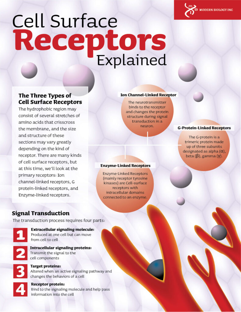
Cell Surface Receptors Explained
Just like people communicate through words, the cells that make up our bodies have a way of communicating to perform the different functions and processes that keep the body alive and responsive.
But of course, there are no talking molecules in the world of cells—they have to use cell receptors and signaling sequences to pass information from one cellular region to another. This is achieved through receptors (receiving molecules) and ligands (signaling molecules).
Receptors and ligands come in various forms, but they always have one trait: they come in pairs, with a receptor recognizing just one (or a few) particular ligands and a ligand attaching to only one (or a few) target receptors. When a ligand binds to a receptor, it alters its shape or function, enabling it to send a signal or cause a change within the cell.
One important component that allows this type of signaling is the cell surface receptor. Let’s look at what exactly cell surface receptors are and their role in converting information from outside the cell into a change within the cell.
In a nutshell, cell surface receptors also known as transmembrane receptors are membrane-linked proteins that bind to ligands (signaling molecules) outside the cell surface and perform signal transduction by converting extracellular signals into intracellular signals.
The ligand doesn’t have to penetrate the plasma membrane in this kind of signaling. As a result, ligands may be a wide range of molecules from large to hydrophilic (water-loving) compounds. A typical cell-surface receptor contains three domains or protein regions:
- An extracellular domain (outside the cell) ligand-binding domain,
- A hydrophobic domain that extends through the membrane
- An intracellular domain (inside the cell) which often sends a signal.

Signal Transduction
A series of biochemical changes that occur inside the cell or the alteration of the cell membrane potential by the flow of ions in and out of the cell sustain signal transmission. Receptors that cause biochemical changes may either directly or indirectly activate intracellular messenger molecules via intrinsic enzymatic activity inside the receptor. The transduction process requires four parts:
- Extracellular signaling molecule: Produced at one cell but can move from cell to cell
- Intracellular signaling proteins: Transmit the signal to the cell components
- Target proteins: Altered when an active signaling pathway and changes the behaviors of a cell
- Receptor protein: Bind to the signaling molecule and help pass information into the cell
The activation and/or inhibition of signaling cascades that control various cellular activities, such as cell growth, proliferation, survival, and invasion, are triggered by ligand binding and cell surface and internal receptors’ activation. The diagram below represents a signal transduction pathway.
The Three Types of Cell Surface Receptors
The hydrophobic region may consist of several stretches of amino acids that crisscross the membrane, and the size and structure of these sections may vary greatly depending on the kind of receptor. There are many kinds of cell surface receptors, but at this time, we’ll look at the three primary receptors: Ion channel-linked receptors, G protein-linked receptors, and Enzyme-linked receptors.
Ion Channel-Linked Receptor
The neurotransmitter binds to the receptor and changes the protein structure during signal transduction in a neuron. This allows extracellular ions to enter the cell by opening the ion channel. As a result, the plasma membrane’s ion permeability is altered, converting the extracellular chemical signal into an intracellular electric signal that affects cell excitability.
The acetylcholine receptor is a cation channel-linked receptor. There are four subunits in the protein: alpha (α), beta (β), gamma (γ), and delta (δ). There are two alpha (α) subunits, each of which has an acetylcholine binding site. Moreover, there are three possible conformations for this receptor. The native protein conformation is closed and unoccupied.
When two acetylcholine molecules attach to the binding sites on alpha (α) subunits, the receptor’s conformation is changed, and the gate is opened, enabling numerous ions and tiny molecules to pass through. However, this open and occupied state only lasts a short time after which the gate is closed, resulting in the closed and occupied state. In this case, the two acetylcholine molecules dissociate from the receptor, restoring it to its original closed and unoccupied condition.
G-Protein-Linked Receptors
The G-protein is a trimeric protein made up of three subunits designated as alpha (α), beta (β), gamma (γ). The alpha (α) subunit releases bound guanosine diphosphate (GDP) in response to receptor activation, which is then displaced by guanosine triphosphate (GTP), resulting in the activating the subunit, which subsequently dissociates from the β, and γ subunits, and the cycle starts all over again. The activated subunit may also have a direct effect on intracellular signaling proteins or target functional proteins.
In eukaryotes, G-protein-linked receptors (GPCRs) are the most numerous and varied category of membrane receptors. Light energy, peptides, lipids, carbohydrates, and proteins all pass via these cell surface receptors, which serve as an inbox for communications. Cells receive these signals to notify them of the presence or absence of life-sustaining light or nutrients in their surroundings or to relay information from other cells.
GPCRs are involved in a wide range of activities in the human body, and a better knowledge of these receptors has had a significant impact on contemporary medicine. In fact, experts believe that GPCRs are involved in the action of one-third to half of all marketed drugs.
Photosensitive chemicals, odors, pheromones, hormones, and neurotransmitters are examples of ligands that bind and activate the G-protein-linked receptors, and they vary in size from small molecules to peptides and large proteins. The cAMP signaling pathway and the phosphatidylinositol signaling pathway are the two main transduction pathways involving G-protein coupled receptors. Both are mediated by the activation of G proteins.
Enzyme-Linked Receptors
Enzyme-Linked Receptors (mainly receptor tyrosine kinases) are Cell-surface receptors with intracellular domains connected to an enzyme. In certain instances, the receptor’s intracellular domain is an enzyme, and in some, the enzyme-linked receptor’s intracellular domain interacts directly with an enzyme. Examples of enzyme-linked receptors include:
- Receptor tyrosine kinases (RTKs),
- Receptor serine/threonine kinases,
- Receptor-like tyrosine phosphatases,
- Histidine kinase-associated receptors, and
- Receptor guanylyl cyclases
Receptor tyrosine kinases are the most common and have the most applications. Growth factors such as epidermal growth factor (EGF), platelet-derived growth factor (PDGF), fibroblast growth factor (FGF), hepatocyte growth factor (HGF), nerve growth factor (NGF), and hormones such as insulin bind to the majority of these molecules.
After binding with their ligands, most of these receptors will dimerize in order to trigger further signal transductions. When the epidermal growth factor (EGF) receptor binds to its ligand EGF, the two receptors dimerize, and the tyrosine residues in the enzyme part of each receptor molecule are phosphorylated. This activates the tyrosine kinase, which then catalyzes further intracellular processes.
Receptor tyrosine kinases can be broken up into seven categories. Each subgroup is represented by just one or two members. In certain subfamilies, the tyrosine kinase domain is interrupted by a “kinase insert region.” The functions of the majority of cysteine-rich, immunoglobulin-like, and fibronectin-type III-like domains are not yet known.
Phosphate groups are removed from phosphotyrosine residues by receptor-tyrosine phosphatases, which counteract the actions of receptor-tyrosine kinases. Receptor tyrosine phosphatases, in many instances, act as negative regulators in cell signaling pathways, ending signals started by protein-tyrosine phosphorylation. On the other hand, some protein-tyrosine phosphatases are cell surface receptors whose enzymatic activities aid cell signaling.
A receptor called CD45, which is expressed on the surface of T and B cells, is an excellent example. CD45 is believed to dephosphorylate a particular phosphotyrosine after antigen stimulation, inhibiting the enzymatic activity of Src family members. As a result, the CD45 receptor-tyrosine phosphatase stimulates nonreceptor protein-tyrosine kinases, which is rather paradoxical.
The extracellular and intracellular domains of enzyme-linked receptors are typically extensive, but the membrane-spanning portion is made up of one alpha-helical peptide strand. When a ligand attaches to the extracellular domain, a signal is sent across the membrane, activating the enzyme, which puts in motion a series of processes inside the cell that ultimately results in a cellular response.
Cell disorders such as cancer are defined by anomalies in cell growth, proliferation, differentiation, survival, migration, and anomalies in signaling through enzyme-linked receptors. This is evidence that Enzyme-linked receptors play a significant role in the development of this class of illnesses.
There are many lab experiments that can be conducted to observe the cell surface structure and help you familiarize yourself with the current strategies used in the study of cell surface receptors and other important regulatory molecules. At Modern Biology Inc, we have high-quality educational products and experiments to help students and tutors when learning about cell surface receptors and other important biological concepts. For more information, reach out to us at (765) 446-4220 or fill out our online form, and one of our team members will get back to you in a jiffy.
 Due to Customs restrictions, we only accept orders from educational institutions within the Continental United States, Alaska or Hawaii.